


In the case of thoracic osteochondrosis, organs associated with the spinal cord region (at the level of the affected thoracic region and below) are often affected. Violation of the normal movement of the spine can lead to immobility of the arms, legs and trunk as a whole, dysfunction of the pelvic organs, respiratory muscles and internal organs.
Osteochondrosis is a degenerative dystrophic disease of the spine that is based on changes in the intervertebral disc, a pathological process involving adjacent vertebrae and intervertebral joints throughout the ligamentous apparatus.
Anatomical features of the spine
The mobility and stability, elasticity and elasticity of the spine are largely determined by the intervertebral disc, which is one of the types of cartilage connections between bones and provides a strong bond between the bodies of adjacent vertebrae. The total length of the intervertebral disc is a quarter of the length of the spine.
The most important function of the intervertebral disc is to reduce the vertical load on the vertebrae. A disk consists of three parts:
- transparent plate (adjacent to the vertebrae);
- nucleus pulposus (fills the gap between the plates);
- Annulus fibrosus (surrounding the nucleus from the outside).
The nucleus contains chondrocytes, tightly wound collagen fibers and chondroitin (proteoglycans). The anterior surface of the disc is covered by the anterior longitudinal ligament, which fuses tightly with the vertebrae and rolls freely over the disc. The posterior longitudinal ligament is firmly fused to the surface of the intervertebral disc to form the anterior wall of the spinal canal. The intervertebral disc does not have its own blood supply, so it feeds on material diffused from the vertebral body.
The vertical load distribution in the spine occurs due to the elastic properties of the intervertebral disc. Due to the pressure, the nucleus pulposus dilates and the pressure is redistributed to the annulus fibrosus and lamina pellucida. During the movement, the core moves in opposite directions: when bending - towards the convex surface, when straightening - forward. When the spine moves, muscles, ligaments, and discs all get involved. Therefore, the violation of one link will lead to the violation of the entire power chain.
Causes and mechanisms of disease development
Mechanical action on the spine plays a special role in the development of osteochondrosis. Under the influence of unfavorable static and dynamic loads, the nucleus pulposus gradually loses its elasticity (as a result of polysaccharide depolymerization), forming protrusions and spacers.
The process of intervertebral disc degeneration is influenced by genetic susceptibility, which leads to changes in the neuromuscular organs of the back, changes in the structure of glycosaminoglycans, and disruption of the distribution of collagen fibers in the intervertebral disc. Genetic factors are crucial in the development of thoracic osteochondrosis and are influenced by functional activity.
Risk factors for the development of spinal degenerative changes include the anatomical features of the intervertebral discs, which are defects in evolution. One of these features is the nutritional profile of the structure. In humans, the intervertebral disc is composed of poorly perfused tissue. Closure of blood vessels occurs already in childhood. After nutrition occurs due to diffusion of substances through the endplates.
The stimulator of nutrient penetration is a dose load that precludes static postures and enormous stress. Inactivity is one of the main risk factors for thoracic osteochondrosis. Therefore, regular exercise is an important preventive measure.
The specificity of the microstructure - a few cells - reduces the strength of the regenerative capacity and the recovery rate of the disk assembly. Anatomical features are weakness and lack of strength in the posterior disc. This contributes to the appearance of wedge-shaped discs in the lower thoracic and lumbar regions.
In the development of osteochondrosis, involutional changes are very important. Active degenerative changes begin to increase after 30 years. Synthesis of the components needed for the disks (glycosaminoglycans) continues, but their quality is deteriorating. The hydrophilicity decreases, the fibrousness increases, and hardening occurs.
Stages of disc degeneration:
- Long-term asymptomatic course, degenerative changes in the components of the disc, displacement of the inner core of the disc;
- Prominent radicular symptoms of osteochondrosis of the thoracic vertebrae, compression of the spinal cord, herniation of the nucleus pulposus (herniation, 1st degree);
- Intervertebral disc rupture with herniated herniation (hernia, degree 2);
- Degenerative changes in the extradiscal component (grade 3).
Pathological protrusions compress nerve roots, blood vessels, or spinal cord at different levels (cervical, thoracic, lumbar), which determine the clinical presentation.
The limited mobility of the thoracic spine, due to the presence of the chest, helps reduce trauma to the intervertebral discs, thereby reducing osteochondrosis. Physiological thoracic kyphosis helps to redistribute the weight of the upper body to the lateral and anterior portions of the vertebrae. As a result, intervertebral hernias and osteophytes form on the front and sides of the spine. Posterior osteophytes and hernias are extremely rare.
Osteochondrosis causes narrowing of the intervertebral foramen and compression of the roots of the spinal cord and sympathetic nerve fibers. Sympathetic nerve fibers originate in the gray matter of the spinal cord and then aggregate into nodes from which they are sent to all internal organs. This results in thoracic osteochondrosis leading to visceral organ dysfunction (vegetative, vasomotor, nutritional) and mimicking somatic disease in addition to typical neurological disorders. This feature of thoracic disc osteochondrosis explains the difficulty of diagnosing and prescribing correct treatment.
Symptoms of thoracic osteochondrosis
Thoracic osteochondrosis is more typical for people with sedentary lifestyles. At the same time, dose loading has no stimulatory effect on the spine, which can lead to interruption of disc recovery. People who work in front of a computer for a long time, bend over, etc. , can get sick. These individuals require independent therapeutic practice.
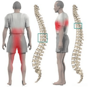
In most cases, thoracic osteochondrosis presents with dull pain, less pain and burning. The pain is located between the shoulder blades. The patient is disturbed by a feeling of pressure in the chest. Localized pain is found when the spinous processes of the thoracic spine are felt, increasing with axial loading of the spine, deep inhalation, and body rotation.
Many patients have severe pain in the shoulder blades and lower chest (posterior rib syndrome). This symptom is the result of displacement of the lower ribs. The pain increases sharply when turning the trunk. More often, the pain syndrome disappears suddenly.
Often chest pain becomes a girdle, corresponding to the process of the intercostal nerves. The sensitivity of the innervated areas of the corresponding nerve endings is disturbed, paresthesia occurs, and there is often a decrease in superficial and deep sensitivity. May violate the function of abdominal compression, alter knee and Achilles tendon reflexes.
Impaired visceral function occurs when any nerve root is compressed at the thoracic level 1 to 12. In the thoracic region, there are structures responsible for innervation of the lungs, heart, intestines, liver, pancreas and kidneys. Therefore, there are no signs specific to thoracic osteochondrosis.
The disease manifests as symptoms of another pathological feature:
- Difficulty breathing;
- severe nighttime pain;
- "heart", angina;
- breast pain;
- right or left rib pain (a symptom of cholecystitis and pancreatitis);
- sore throat and esophagus;
- upper abdominal pain (symptoms of gastritis, enteritis, and colitis);
- Sexual dysfunction.
diagnosis
The greatest value in diagnosing thoracic osteochondrosis is an X-ray of the chest. The picture shows decreased disc height, endplate sclerosis, and osteophyte formation.
Computed tomography scans allow you to determine the condition of the vertebrae, the joints of the spine, the size of the spinal canal, and the location and size of herniated hernias.
When making a differential diagnosis, it is necessary to carefully collect the medical history and compare all the clinical symptoms of sternal osteochondrosis with those of other diseases. For example: heart pain in osteochondrosis cannot be stopped by nitroglycerin, epigastric pain is not related to food intake, not seasonal, all symptoms appear mainly at night and disappear completely after a night of rest.
How is thoracic osteochondrosis treated?
Treatment of thoracic osteochondrosis is conservative in almost all cases. The indication for treatment is the predominance of visceral syndromes with neurological disorders. The primary orthopaedic treatment should be adequate spinal traction:
- Underwater active vertical traction;
- Passive horizontal traction using a Glisson ring on an inclined bed was used in cases of impairment at the level of 1-4 thoracic vertebrae, and traction was performed using axillary girdle in cases of impairment at the level of 4-12 thoracic vertebrae.
Drug therapy includes paravertebral block with neocaine solution. Analgesics and sedatives are used as the disease worsens. For unexpressed pain syndromes, ointments containing analgesics and anti-inflammatory drugs are permitted at home.
Use the back and lower extremity muscles to massage after the acute symptoms are resolved. In cases of functional blockade, manual therapy is indicated for osteochondrosis of grades 1-3. It includes various options for soft and rough effects on the back muscles.
Therapeutic exercises allow you to load all parts of the spine in a dosed manner that facilitates the recovery process. An important condition for exercise therapy in osteochondrosis is the exclusion of vertical loads.
Physiotherapy: UHF therapy, ultrasound, induction heat therapy, radon and pine needle salt baths. During the spa phase, actively use underwater traction and hydromassage.
Surgery is rarely used. The indication for surgical intervention is compression of the spinal cord by fragments of the prolapsed disc.